
Medical Imaging
Adventist Health Mendocino Coast’s diagnostic imaging services combines state-of-the-art technology with convenience and comfort. Diagnostic services are conveniently located at the hospital and staffed by highly trained and compassionate technologists with board-certified radiologists interpreting the results.
To learn more or to schedule an appointment, call (707) 961-4665.
3D mammography
3D mammography is proven to provide greater accuracy detecting breast cancer. By taking multiple pictures of each breast in just four seconds, a 3D mammogram gives radiologists clear, highly focused images of the breast’s tissue, layer by layer. 3D mammography increases detection of all breast cancers by 29 percent and increases detection of invasive breast cancers by 41 percent. 3D mammography results in fewer unnecessary biopsies and reduces call backs by 41 percent.
Bone densitometry (DEXA) scan
DEXA or bone densitometry examinations estimate the amount of bone mineral content in specific areas of your body. Two X-ray energies allow the radiologist to tell the difference between bone and soft tissue, giving a very accurate estimation of your bone density.
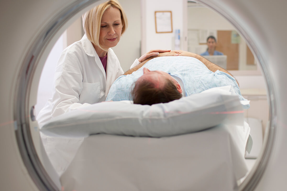
Cardiac CT scan
In a cardiac CT scan, a state-of-the-art volume computed tomography (VCT) scanner creates 64 paper-thin, cross-sectional images of the resting heart in a matter of seconds. These images are then assembled into a complex, three-dimensional picture of the heart and its arteries. The scan provides extraordinary detail and can detect cardiac abnormalities that lead to treatment if necessary.
Computed tomography CT scanning
CT scanning is an imaging technology that uses multiple channels and computers to create detailed, cross-sectional images of specific areas of the body. These images may be used to identify fractures, infections or tumors.
A CT scan is frequently used to evaluate abnormalities like blocked blood vessels. CT can serve as a valuable tool for use in minimally invasive interventional procedures. Ukiah Valley Medical Center imaging strives to keep radiation doses as low as possible.
Digital x-ray
X-rays are a form of electromagnetic radiation, just like visible light. X-ray machines send individual X-ray particles, called photons, which pass through the body and are recorded by computer to create digital images.
Magnetic resonance imaging (MRI)
MRI is an advanced imaging technique that uses a high-field magnet to produce high-resolution digital images of soft tissue and structural anatomy. MRI has revolutionized the fields of musculoskeletal, cardiovascular and neurologic imaging.
Nuclear medicine imaging
Nuclear medicine uses radiopharmaceuticals to diagnose a variety of diseases. It can be used to evaluate inflammatory conditions and is useful in detecting some cancers.
Open Magnetic Resonance Imaging (MRI)
An open MRI has a larger opening then a standard MRI. This is beneficial for those that get nervous in small places (claustrophobic) and for people who are physically large.
Ultrasound or sonogram
Ultrasound uses high-frequency sound waves to capture diagnostic cross-sectional images of the targeted part of the body, typically used for imaging abdominal and pelvic organs, prenatal testing, blood vessels and small parts (like breasts, thyroid and testicles). A doctor’s order is required.